What’s been happening in 2018/19?

Picture1
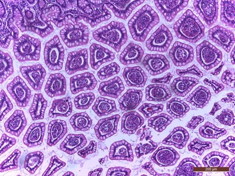
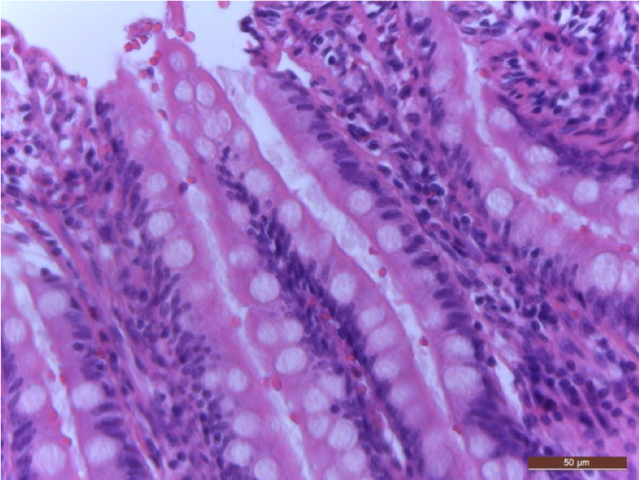
Oh Goblet cells, oh goblet cells……………..

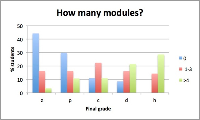
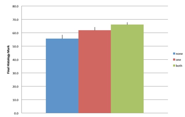
In 2017/2018 Histology students created over 100 new images and MCQ for HistoLab. Two students spent their science internship over the summer holidays rejuvenating and upgrading the HistoLab portal to include 16 modules.

My name is Rana; I am an undergraduate student who finished Histology in 2018. During my histology studies, I was glad that we had such an excellent study resource “HistoLab modules” which made studying histology easier and helped me a lot during practical classes. For those reasons, my colleagues Ingrid and I spent our summer holidays updating and generating new Histolab modules for 2019 Histology students using the stored image banks of 2017 & 2018 student-generated MCQ and images. We had a lot of fun and a remarkable three weeks recreating these modules. We generated a module for every topic using images which was taken from slides that will be used during practical classes. I hope you guys appreciate this valuable study recourse and I encourage you all to use it before practical classes for easier identification of histological structure.  Have fun and enjoy studying such an interesting subject with awesome teaching staff.

My name is Ingrid, and together with Rana I have been updating the Histolab modules. I hope you will find them useful and use them, as I think they are a great learning resource to use before you come to your practical class. As a former histology student I would also advise that you look at the additional learning resources (my favourite was University of Michigan “Webscope”)


What students said in 2017

Picture1

This is one of the fantastic images taken by a histology student in 2017. Do you know what it is? These are some of the comments given by students as feedback about HistoLab! I look forward to hearing what you think.

i will know what the prac class comprises of.

The more resources and questions, the better my knowledge and understanding plus retention will be.

a useful tool to assess where I’m at and how much more work i need to put back into the topic.

Allows for problems to be identified before practicals and after lectures therefore allowing me to ask questions and evaluate my learning.

Definately has been really helpful for me and reinforced todays lecture.

I loved this module, it forced me to research and I learned a lot from completing this.

This just reinforces the knowledge from the lecture and feels like a practice run for the sorts of questions I might expect in a formal exam for this subject.

This was a good indicator for what to expect in the practicals as well as practice for identification

this was a massive help as it showed me how much i really know on the top of my head


HistoLab Going strong

Students in 2015 and 2016 have generated over 200 images to include in new HistoLab modules. We have expanded from 5 to 10 modules in 2017, and these ones have been mostly written by finishing students so will be good preparation for new students. Some of them are pretty hard, and will be quite challenging. HistoLab is offered as voluntary pre-lab activities using microscope images from the class teaching sets. Where ever possible feedback will be given to those students who have completed them, and they will be available for revision for everyone.

Students who have completed HistoLab Modules before coming to class continue to out-perform those that didn’t do them. This graph shows that approximately 70% of all the students who completed 4 or more modules passed this subject including 30% who got high distinctions, compared to those who did less than 4 modules (~55% and 15% HD) or didn’t do any (~48% and no HD). 5-modules

Growing HistoLab

In 2015, histology students at UTS will have the opportunity to be actively involved in growing HistoLab. During their practical classes they can select a slide from the teaching set, take a digital image and write a multiple choice question.

This gives students the chance to experience new microscopy techniques, learn how digital imaging software is used, participate in learning and teaching activities with their peers and learn how to work together to create an interactive learning space.

The best of the images and questions will be used to make new Histolab modules, and may even pop up in assessment tasks. Students designing for students! What better way to learn what you are doing than by teaching someone else?

Did HistoLab help students?

Did HistoLab help students?

This is a graph showing the final grades for students in HIstology 2013. The average grade was about 55. Students that participated in HistoLab got, on average, 5-10 more marks than students that didn’t do either module. This was a statistically significant difference.

Students that did any HistoLab module were also more likely to get a credit grade or higher than students that didn’t participate.

So…….HistoLab certainly appears to be helping students with their studies. Whether this is through assisting with lab preparation, reinforcing key concepts or guiding revision, the overall effects are very positive for student learning.

2nd Module

2nd Module

…and then there was stomach. This module was run through the second part of the semester, and was much more complicated than module 1. Students needed to examine low and high power images of the different parts of the stomach, and think about how function relates to structure.

It was especially aimed at getting students to recognise the different layers in the organs wall, how these layers are common to many parts of the gastrointestinal tract, and where they differ. Specialised cells and their function in digestion were also visited.

Not as many students completed this module, which was a shame. However, comments from completing students were still very positive. Image quality was improved and was appreciated.

1st Module

1st Module

The first module made available to students was all about muscles. Cardiac muscle, skeletal muscle and smooth muscle [what type is shown here?]. There were three images of each showing three identifying features. After the lecture, students had a week to go on-line to examine the images and answer questions before attending the practical class. The answers were posted that morning and I could see which concepts were less well understood, and go through these in a little more detail during the class.

What did the students think?
Over half the enrolled students completed this module. It only took 10-20 minutes to do and 80% of those said they would benefit from doing more modules.

This is what they had to say………….

– Very useful. Re-enforces content in a way which makes learning fun
– This was actually a really useful feature to do before the prac, thanks. would be really great to keep these up for revision, as its much better to learn with access to reliable images and slides. Thanks.
– i found this helpful ! even questions i was unsure about i could look bak to my notes and rein-enforce lecture material.
– Really great system to help students in learning the features of muscles cells, this will definitely help me in preparing what to look for as of tomorrow’s laboratory class as i am always unsure of what to look for during pracs
– Very nice to have annotated digital images outside of pracs.
– i think it helped to have the digital picture available outside of class and it helped reinforce the structure
– Make more of these modules

BUT we need to work on the image quality……..
– Some were a bit difficult to see at their current size.
– images are too small and appear blurry when I zoom in
– Should have a zoom feature in order to view the images at a closer level
– Higher resolution images. The images were of such bad quality
– The images didn’t have high enough resolution, so it was difficult to pick out fine details

Soon, we will assess whether students that completed this module preformed better in their assessment tasks than those that didn’t.

What is HistoLab?

HistoLab

HistoLab is a portal designed for students studying Histology at the University of Technology, Sydney. It gives students an opportunity to access digital images of microscope slides before they come to laboratory class. Students view digital images of histology sections and answer a series of multiple choice questions aimed at reinforcing the threshold concepts necessary to understand this subject. They will receive feedback showing their answer, the correct answer, and the class answer. Importantly, I can also see how the entire class has responded, which allows me to identify gaps in the knowledge and concentrate on specific areas during class time.

Each module covers aspects of one topic in the curriculum. A module takes about 10 minutes to complete. HistoLab modules are not compulsory, nor are they assessed, but it will benefit students to complete these modules, to refer to their textbook and lecture notes, their practical manuals and do some independent research as they go. Then, when they attend practical class and have hands on access to the slides and microscopes, they will know what to look for and where. They will be able to use their time more efficiently in class investigating the more detailed morphology and specializations of individual cells and tissues.

Histology images available in textbooks tend to be “gold standard”. Histology slides in real life can be far from perfect. Our teaching slides are unique. HistoLab gives students the opportunity to have a sneak peek before they come to class. HistoLab can be used a pre-lab tutorial, a quiz or as a revision tool. Students can complete modules in their own space and in their own time and they can come back and try again later.